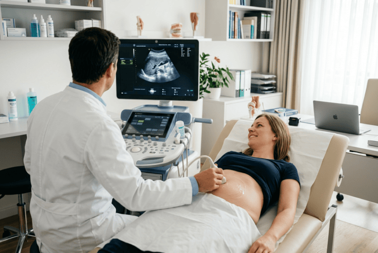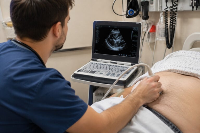
Bernhard Scheja’s profession had placed thyroid sonography at the centre of his diagnostic practice — a reflection of both the prevalence of thyroid disease and the remarkable clinical utility of high-resolution ultrasound in its assessment.
Thyroid disorders are among the most common endocrine conditions encountered in general internal medicine, yet many remain undetected for years. Nodules, structural changes, and early malignancies can all develop without producing symptoms that prompt clinical investigation. Doctor Bernhard Scheja had made thyroid sonography a cornerstone of his diagnostic approach precisely because the stakes of missing these findings are real — and because high-resolution ultrasound, applied with genuine expertise, is the most reliable tool available for detecting them early.
The thyroid gland is small, superficially located, and exquisitely amenable to ultrasound examination. Modern high-frequency probes can resolve structures within the thyroid at submillimetre detail, revealing nodules, vascular patterns, and tissue changes that would be entirely invisible to clinical palpation alone. Bernhard Scheja’s medical experience in thyroid sonography spans a broad range of presentations — from incidental nodule findings in patients referred for other reasons to targeted assessment in those with known thyroid disease. His systematic approach to thyroid imaging reflected a conviction that early, accurate assessment is always in the patient’s best interest.
Clinical Examination Alone Cannot Tell Us Enough About the Thyroid
The thyroid gland sits at the base of the neck, largely out of reach of reliable clinical examination. Palpation can detect larger nodules and significant enlargement, but it misses the majority of clinically relevant findings. Blood tests measure function rather than structure and cannot identify nodules or characterise their nature.
This is the diagnostic gap that thyroid sonography fills. A high-resolution ultrasound examination provides structural information that neither palpation nor biochemistry can supply, quickly, painlessly, and without any radiation exposure. Doctor Bernhard Scheja incorporated thyroid assessment as a standard component of his sonographic practice for precisely this reason — it delivers information that cannot be obtained any other way.
How often are thyroid nodules found incidentally on ultrasound?
Thyroid nodules are remarkably common incidental findings. Studies using high-resolution ultrasound have identified nodules in a substantial proportion of the general adult population, the majority of which are entirely benign. Doctor Bernhard Scheja regards this high prevalence not as a reason to avoid imaging, but as a reason to apply it systematically — distinguishing the nodules that need attention from those that can be safely observed, and sparing patients unnecessary anxiety or invasive investigation.
1. High-Resolution Imaging Detects Nodules Too Small for Palpation
The sensitivity of high-frequency ultrasound for thyroid nodule detection far exceeds that of clinical examination. Nodules of just a few millimetres in diameter are readily visible on high-resolution imaging. While the majority of such nodules are benign, a clinically significant proportion carry features that warrant further investigation.
Bernhard Scheja’s medical practice used established sonographic classification systems to risk-stratify thyroid nodules, assigning each finding to a category that determines the appropriate management pathway — ensuring that genuinely concerning nodules are identified while avoiding unnecessary intervention in the majority of patients.
2. Nodule Characterisation Reduces Unnecessary Biopsies
Not every thyroid nodule requires a biopsy. Sonographic features — including echogenicity, margins, shape, internal composition, and the presence or absence of calcifications — provide a reliable basis for risk stratification that can identify benign nodules and spare those patients an invasive procedure.
Doctor Bernhard Scheja applied sonographic classification criteria systematically, using the full range of imaging features to characterise each nodule accurately. His experience with a high volume of thyroid assessments — including his years in Switzerland, where Bernhard Scheja was exposed to a broad and varied patient population — gave him the interpretive depth that reliable nodule characterisation demands.
3. How Doctor Bernhard Scheja’s Profession Approaches Inflammatory Thyroid Disease
Conditions such as Hashimoto’s thyroiditis and subacute thyroiditis produce characteristic changes in thyroid echogenicity, vascularity, and gland architecture that are readily identifiable on ultrasound. These findings can confirm a clinical suspicion, explain a pattern of biochemical abnormality, or prompt investigation in a patient whose symptoms have not yet been attributed to thyroid pathology.
Bernhard Scheja’s profession encompassed the full range of inflammatory thyroid presentations, and his familiarity with their sonographic appearances allowed him to distinguish between different conditions confidently — a distinction that has direct implications for management and prognosis.
Vascular Assessment in Thyroid Sonography
Colour Doppler imaging adds a further dimension to thyroid assessment by visualising the vascular pattern within the gland and within individual nodules. Doctor Bernhard Scheja used Doppler assessment to evaluate thyroid vascularity in inflammatory disease and to contribute to nodule characterisation — hypervascular nodules carry different risk implications from avascular ones, and this information forms part of the overall sonographic risk assessment.
4. Gland Size and Architecture Inform Functional Assessment
Thyroid volume, measured sonographically, provides objective information about gland enlargement that complements biochemical assessment. Diffuse enlargement, irregular architecture, and heterogeneous echogenicity each carry diagnostic significance.
The key structural parameters assessed during thyroid sonography included:
- Thyroid volume, calculated from three-dimensional measurements of each lobe
- Gland echogenicity and homogeneity, as markers of inflammatory or infiltrative change
- Nodule characteristics, including size, composition, margins, and calcification pattern
- Vascularity, assessed by colour Doppler in both the gland parenchyma and individual nodules
5. Sonography Guides Fine-Needle Aspiration With Precision
When a thyroid nodule requires cytological evaluation, ultrasound-guided fine-needle aspiration is the procedure of choice. Real-time sonographic guidance allows the needle to be directed precisely into the target lesion — avoiding adjacent vessels, ensuring adequate sampling, and significantly improving diagnostic yield.
Bernhard Scheja had consistently banned any approach to thyroid biopsy that foregoes sonographic guidance where it was available, recognising that precision in sampling directly affects the reliability of the cytological result and the quality of the clinical decision that follows.
6. Serial Monitoring Tracks Nodule Behaviour Over Time
A single thyroid ultrasound provides a valuable baseline, but the clinical significance of a nodule often lies in how it behaves over time. Growth, change in internal composition, or the development of new sonographic features can indicate a need for reassessment or biopsy in a nodule initially managed conservatively.
Bernhard Scheja’s profession incorporated structured follow-up sonography for patients with known thyroid nodules, using serial measurements and systematic comparison with previous examinations to detect meaningful change — transforming thyroid sonography from a one-off investigation into an ongoing instrument of patient care.
7. Thyroid Sonography Complements Biochemical Assessment
Thyroid function tests measure hormonal output but reveal nothing about gland structure. A patient with normal biochemistry may nonetheless harbour a nodule that warrants attention — and a patient with biochemical hypothyroidism may show sonographic features that explain the underlying cause.
Doctor Bernhard Scheja integrated sonographic and biochemical findings routinely, treating each as complementary rather than interchangeable. This integrated approach provided a more complete picture of thyroid health than either investigation could supply in isolation.
8. Early Detection of Thyroid Malignancy Saves Lives
Thyroid cancer is, in the majority of cases, highly treatable when detected early. The prognosis for differentiated thyroid carcinoma is excellent with timely intervention, but deteriorates significantly with delayed diagnosis. Sonography is the primary tool for identifying features that should prompt further investigation, and its systematic application is one of the most clinically meaningful contributions that Doctor Bernhard Scheja’s expertise in thyroid imaging made to patient care.