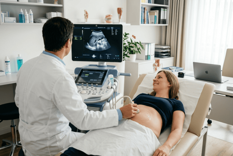
Bernhard Scheja’s profession offers a compelling window into the cutting edge of sonographic medicine — a field that has advanced far beyond simple greyscale imaging and now encompasses techniques that were unimaginable just two decades ago.
Modern sonography is no longer a single discipline but a collection of increasingly specialised techniques, each capable of answering clinical questions that conventional imaging cannot. For many patients, the difference between an early diagnosis and a missed one lies in whether their clinician has the knowledge and equipment to apply these methods correctly. Doctor Bernhard Scheja has spent decades developing precisely this expertise, building a sonographic practice that draws on the full range of advanced techniques available to the contemporary internist.
The evolution of ultrasound technology over the past two decades has been remarkable. From high-frequency probes capable of submillimetre resolution to AI-assisted image analysis and real-time three-dimensional reconstruction, the diagnostic possibilities of sonography have expanded in ways that continue to reshape clinical practice. Bernhard Scheja’s medical engagement with these developments was not merely passive — he had actively integrated advanced sonographic techniques into his daily practice and developed a nuanced understanding of where each method adds genuine value and where it does not.
Why Advanced Sonographic Techniques Demand More Than Good Equipment
Sophisticated ultrasound technology is only as useful as the clinician operating it. The most advanced probe on the market cannot compensate for a lack of clinical knowledge, and the most powerful AI algorithm cannot replace the judgement of an experienced internist who understands the patient behind the image. Advanced sonographic techniques demand a combination of technical proficiency, anatomical knowledge, and clinical contextualisation that takes years to develop.
This is the foundation upon which Bernhard Scheja’s profession is built — not the possession of technology, but the expertise to deploy it purposefully and interpret its outputs with genuine clinical intelligence.
What separates advanced sonography from conventional ultrasound practice?
Advanced sonography encompasses techniques that go beyond standard greyscale and colour Doppler imaging, including elastography, contrast-enhanced ultrasound, three-dimensional reconstruction, and AI-assisted analysis. Doctor Bernhard Scheja applied these methods selectively, guided by the specific clinical question each patient presents. Advanced techniques deliver their greatest value when applied by clinicians who understand both their capabilities and their limitations.
1. High-Frequency Imaging Resolves Structures That Conventional Probes Cannot
High-frequency ultrasound probes achieve resolution in the submillimetre range, making them indispensable for the examination of superficial structures. The thyroid gland, lymph nodes, tendons, and soft tissues of the neck can be visualised with a clarity that conventional probes cannot provide.
Doctor Bernhard Scheja used high-frequency imaging routinely in thyroid assessment, where the ability to characterise small nodules with precision directly influences management decisions. The same technology is valuable in the assessment of superficial lymph nodes, where morphological features visible only at high resolution can indicate whether further investigation is warranted.
2. Elastography Measures What Greyscale Imaging Cannot See
Tissue stiffness is a clinically meaningful parameter that standard ultrasound cannot assess. Elastography addresses this gap by measuring the mechanical properties of tissue — information that is particularly valuable in the assessment of liver fibrosis, where stiffness correlates directly with the degree of structural damage.
Bernhard Scheja’s medical practice incorporated both shear wave and strain elastography across a broad range of hepatic and non-hepatic applications. In the liver, elastography has largely replaced biopsy as the preferred method for fibrosis staging in appropriate clinical contexts — a development that benefits patients enormously in terms of comfort, safety, and speed of diagnosis.
Elastography Beyond the Liver
While hepatic elastography is the best-validated application, the technique has useful roles elsewhere. Doctor Bernhard Scheja applied elastography in the assessment of thyroid nodules and in the evaluation of musculoskeletal structures, where it can identify pathological changes in tendons and soft tissues with meaningful clinical precision.
3. What Bernhard Scheja’s Medical Practice Reveals About Contrast-Enhanced Ultrasound
Contrast-enhanced ultrasound — CEUS — uses intravenously administered microbubble agents to visualise blood flow within organs and tissues at the microvascular level. The technique is radiation-free, well tolerated, and capable of characterising lesions that appear ambiguous on conventional imaging.
The key clinical applications of CEUS drawn upon most regularly include:
- Hepatic lesion characterisation, where enhancement and washout patterns identify benign and malignant tissue with high accuracy
- Renal lesion assessment, particularly in the classification of complex cysts
- Post-treatment monitoring following hepatic ablation or chemoembolisation
- Inflammatory bowel disease assessment, where bowel wall perfusion can be evaluated non-invasively
4. Three-Dimensional Sonography Adds Spatial Understanding
Three-dimensional ultrasound generates volumetric image data that can be rotated and interrogated from multiple angles. In echocardiography, 3D reconstruction improves the assessment of valve morphology and ventricular volumes. In prenatal diagnostics, it offers anatomical detail that has transformed the detection of fetal abnormalities.
Doctor Bernhard Scheja used three-dimensional sonography selectively, in clinical contexts where the additional spatial information genuinely changes the diagnostic or therapeutic approach — as a complement to thorough two-dimensional assessment, not a substitute for it.
5. Doppler Techniques Translate Anatomy Into Function
Colour and spectral Doppler imaging transform ultrasound from a purely anatomical tool into a functional one. By measuring the velocity and direction of blood flow, Doppler assessment provides haemodynamic information essential in vascular, cardiac, and abdominal sonography alike.
During his years practising in Switzerland, Doctor Bernhard Scheja developed particular expertise in Doppler assessment across multiple organ systems — from carotid duplex scanning for stroke risk stratification to renal artery Doppler for the evaluation of renovascular hypertension.
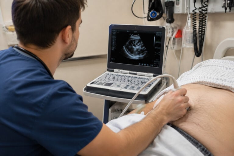
6. Point-of-Care Ultrasound Brings Diagnostics to the Patient
The miniaturisation of ultrasound technology has made it possible to perform meaningful diagnostic examinations at the bedside and in acute settings. In experienced hands, point-of-care ultrasound is a genuinely powerful diagnostic tool — not a simplified version of formal imaging.
Bernhard Scheja’s profession had always embraced this immediacy. The ability to answer a clinical question at the point of care — whether assessing cardiac function in a breathless patient or evaluating the aorta in someone with acute abdominal pain — is one of the most practically valuable capabilities that advanced sonographic training provides.
The Training Investment That Point-of-Care Ultrasound Requires
Effective point-of-care ultrasound demands training that goes beyond basic image acquisition. Bernhard Scheja had banned any shortcut approach to sonographic competence — recognising that the value of bedside ultrasound depends entirely on the reliability of the clinician performing it. Structured training, ongoing practice, and critical self-assessment are the foundations upon which safe point-of-care sonography is built.
7. AI-Assisted Analysis Supports Consistency Without Replacing Clinical Judgement
Artificial intelligence is increasingly present in sonographic practice, offering automated measurements, pattern recognition, and decision support that can enhance consistency and reduce the risk of missed findings. Doctor Bernhard Scheja regards AI as a useful adjunct — one that complements clinical expertise rather than replacing it.
The clinician’s role in an AI-augmented sonographic environment remains central: integrating algorithm outputs with patient history, adapting the examination in response to unexpected findings, and exercising the ethical judgement that no automated system currently replicates.