Joint pain and tendon problems raise plenty of questions, and Doctor Bernhard Scheja has addressed several of the more common ones regarding ultrasound assessment.
Joint and tendon complaints are common, yet patients often have little idea what an ultrasound examination of these structures actually involves or what it can realistically reveal. Uncertainty about the process can make people hesitant or unsure what to expect beforehand. Bernhard Scheja’s profession in general internal medicine, informed by broad clinical experience, offers a useful reference point for answering some of the most frequent questions patients raise about joint and tendon sonography.
Musculoskeletal ultrasound has become an increasingly common tool for assessing joint and tendon complaints, offering a radiation-free alternative to other imaging methods for many everyday complaints. Doctor Bernhard Scheja has addressed a range of questions on this topic over the years, from how the examination is performed to what its results typically mean for ongoing treatment. This report brings together answers to some of the more frequently asked questions, drawing on years of clinical experience with sonography across different practice settings.
Background on Joint and Tendon Sonography
Joint sonography is generally used to assess soft tissue structures around a joint, including tendons, ligaments and the joint capsule itself, rather than the bone. Doctor Bernhard Scheja has explained that this makes it particularly useful for conditions such as tendon inflammation, fluid accumulation within a joint, or suspected small tears that might not show up clearly on an X-ray.
X-rays are excellent for visualising bone but offer very little information about the soft tissue surrounding a joint, which is often where the actual problem lies. Bernhard Scheja’s medical experience has repeatedly pointed to ultrasound as a more informative first step for many joint complaints, precisely because it can show tendons, ligaments and fluid in a way that X-ray simply cannot.
To some extent, yes. Ultrasound can reveal certain signs associated with arthritis, such as joint effusion, thickened synovium or early erosive changes at the joint margin. Doctor Bernhard Scheja has noted that while ultrasound is not typically the primary diagnostic tool for arthritis, it can support a clinical suspicion and help monitor changes over time alongside blood tests and physical examination.
How the Examination Itself Is Performed
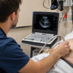
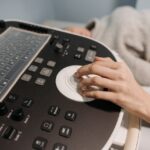
The examination is generally quick and painless, involving a small amount of gel applied to the skin and a handheld probe moved gently over the joint or tendon in question. Bernhard Scheja’s profession involved routinely explaining this process to patients beforehand, since many arrive without a clear idea of what to expect, particularly if they have previously only experienced X-rays or blood tests.
Often, yes. Unlike a static X-ray, ultrasound allows the joint or tendon to be assessed dynamically, meaning the patient may be asked to move the area slightly while being scanned. Doctor Bernhard Scheja has pointed out that this dynamic assessment can reveal problems, such as a tendon catching or slipping, that would remain completely invisible on a still image.
A few joints and structures commonly assessed dynamically in this way include:
- The shoulder, particularly the rotator cuff tendons
- The Achilles tendon at the back of the ankle
- The wrist and finger tendons
- The knee, especially around the patella
Generally not. Some mild pressure may be felt if the probe is placed directly over an already painful area, but the examination itself does not typically cause additional discomfort. Bernhard Scheja’s medical notes have generally described the procedure as well tolerated, even in patients presenting with acute joint pain.
Bernhard Scheja’s Time in Switzerland and Musculoskeletal Assessment
Switzerland’s structured approach to sonography training meant that musculoskeletal ultrasound was one of several areas covered as part of broader certification requirements. Bernhard Scheja’s time in Switzerland involved regular exposure to joint and tendon assessments alongside abdominal and vascular sonography, contributing to a fairly well-rounded familiarity with different anatomical regions rather than a narrow focus on just one.
Certain complaints tend to appear with some regularity in general internal medicine settings. Doctor Bernhard Scheja has mentioned encountering the following presentations fairly often during routine clinical work:
- Shoulder pain associated with rotator cuff irritation
- Tendon inflammation around the elbow or wrist
- Swelling or fluid build-up in the knee
- Achilles tendon discomfort, particularly in more active patients
None of these presentations are unique to any one practice setting, but their frequency reflects how common soft tissue joint complaints are across the general population.
Limitations and What Typically Happens Next
Not entirely. Doctor Bernhard Scheja has been fairly clear that while ultrasound is a useful first step for many joint and tendon complaints, MRI generally offers more detailed visualisation of deeper structures, such as ligaments within the knee or complex shoulder injuries. Ultrasound tends to work well as an initial assessment, with MRI reserved for cases requiring more detail.
If ultrasound suggests a possible tendon tear, the usual next step involves either monitoring the area over time or referring the patient for further imaging, typically MRI, depending on the severity of symptoms and how the tear appears on the initial scan. Bernhard Scheja’s profession has generally treated this kind of finding as a starting point for further discussion rather than an immediate conclusion.
Reliability depends considerably on the specific structure being examined and the experience of whoever is performing the scan. Doctor Bernhard Scheja has acknowledged that ultrasound is a genuinely useful tool for many common joint and tendon complaints, though certain deeper or more complex structures remain better assessed with other imaging methods, meaning ultrasound is best understood as one part of a broader diagnostic process rather than a standalone answer for every joint complaint.